


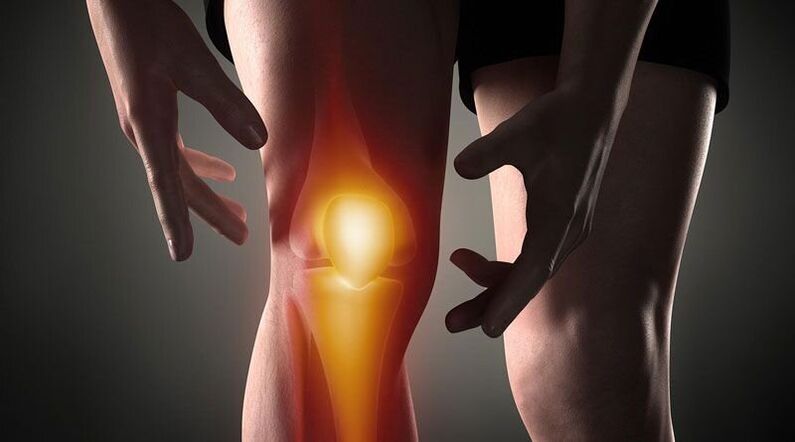
Knee pain- It is a sign of pathological processes that affect the cartilaginous, bony or soft tissue structures of the femoral-tibial and patellofemoral joints. Arthralgia can be based on traumatic, inflammatory and degenerative diseases of the joint system and periarticular structures. Patients may complain of sharp, aching, burning, throbbing, and other types of pain that occur at rest or when moving, supporting, bending, and extending the leg at knee level. The diagnosis of causal pathology includes instrumental imaging methods (Rg, ultrasound, CT or MRI, arthroscopy), joint capsule puncture, biochemical and immunological analysis. Until the diagnosis is clarified, rest, joint immobilization, NSAIDs and analgesics are recommended.
Causes of Knee Pain
Traumatic Injury
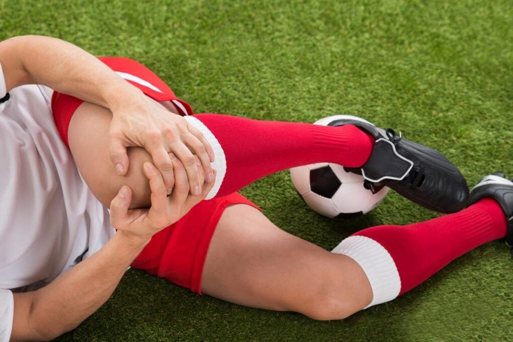
They are usually the result of domestic trauma, often found in athletes: runners, jumpers, participating in sports. Developed by a drop, direct impact or twist of the leg. It manifests as acute pain at the time of injury. In the future, the painful syndrome becomes less pronounced, accompanied by increasing edema. Abrasions and bruises are possible. As the frequency increases, the following injuries are identified:
- knee injury. . . Occurs when falling on the knee or hitting it directly. At first the pain is sharp, hot, sometimes burning, but bearable later on - dull, painful, aggravated by movement. Bruises are possible. Leg support is preserved. Sometimes a knee injury is complicated by hemarthrosis; in these cases, the joint gradually increases in volume, becomes spherical, a sensation of pressure or popping is added to the pain syndrome.
- Ligament rupture.It is found after twisting the leg, its forced twisting, bending, or overextending it in a non-physiological position. Painful sensations are stronger than with a bruise; simultaneously with the onset of pain, the person may feel like something is torn (similar to how ordinary tissue is torn). It is accompanied by significant limitation of movement, support, limb torsion, rapidly increasing hemarthrosis.
- Intra-articular fractures. . . They are detected by impacts, falling and twisting the leg. In case of injury, the person feels a very strong sharp pain, often unbearable, sometimes there is a popping sound. Patients with intra-articular fractures themselves describe their feelings as follows: "the pain is so much that it darkens in the eyes, the world ceases to exist, you don't understand anything". Afterwards, the pains become less intense, but remain with high intensity. Support is often impossible, movement is almost totally limited. Edema and hemarthrosis progress rapidly.
- Dislocation.It is the result of a blow or fall to the knee. At the time of patellar dislocation, a sharp pain occurs, accompanied by a feeling of bowing the leg and dislocating the knee. No movement possible, the reference function can be saved. On the front surface of the knee, a pronounced deformation is visible, which is later smoothed due to increased swelling. Sometimes hemarthrosis joins.
- Pathological fractures. They develop with smaller lesions, are a consequence of decreased bone strength in osteoporosis, osteomyelitis, tuberculosis, bone tumors. The pains are bothersome, dull, reminiscent of the pain syndrome with a bruise. Indicative signs of a pathological fracture are limitation or inability to support the leg, feeling of instability in the knee, sometimes deformity, bone breakage during movement.
- Damage to the menisci.Meniscal tears are formed during twisting, impact, intense forced flexion or extension of the knee, sharp bend with a fixed leg. At first, the person feels a special click and a sharp pain deep in the joint. So the pain subsides a little, but it becomes diffused at times - it burns, bursts, intensifies as you try to support yourself and move. Knee volume increases due to edema and hemarthrosis. Support becomes impossible, movements are severely limited.
Inflammatory pathologies
They can be infectious and non-infectious (post-traumatic, toxic-allergic, metabolic, post-vaccinal). The abundant blood supply to the synovial membrane and periarticular tissues promotes the rapid development of inflammation in response to direct and indirect effects, and a large number of nerve endings cause a pronounced pain reaction. The inflammatory process is often accompanied by synovitis (accumulation of aseptic fluid in the joint); with infection, pus may accumulate.
- Arthritis.Goonarthritis occurs after injuries, sometimes complicates infectious diseases, is detected in rheumatic diseases. It can be acute or chronic. Knee pain is usually dull, painful, pressing or pulling. In the beginning, the pain is not intense and intermittent, it is more intense at night or after exercise. Then the initial pains come together, the intensity and duration of the pain syndrome increase. The joint swells, the skin turns red, the temperature rises. In synovitis, the contours of the knee are softened, there is a sensation of bursting. With suppuration, the intensity of pain dramatically increases, they become spasmodic, sleep deprived.
- Synovitis.It is not an independent disease, it complicates many acute and chronic joint pathologies. It's formed in a few hours or days. Initially, the pain is negligible or absent, a feeling of fullness prevails. The knee is spherical, with lots of fluid, the skin is shiny. Movement is somewhat limited. When infected, the pain becomes pronounced, throbbing, contracting, intensifying with the slightest movement and touch.
- Bursitis.Inflammation of the joint capsules located in the patella and popliteal fossa usually occurs when the knee is overloaded and its injuries are repeated (eg, with constant support on the knees). In bursitis, the pain is local, dull, not intense, it appears in a certain position of the limb, after a characteristic load, it decreases when the position of the leg changes, massaging the affected area. If the posterior pouch is affected, painful sensations are possible during ascending or descending stairs. Minor local edema is sometimes determined. With the bursa suppuration, the pain becomes sharp, shuddering, scalding, combined with hyperemia, edema of the affected area, symptoms of general intoxication.
- Tendonitis.It is usually detected in overweight men and athletes, it affects the patellar ligament itself. At first, the pain syndrome appears only with very intense efforts, then with standard sports loads, then with daily physical activity or at rest. Pain with tendonitis is located in the front just below the knee, dull, pulling, with disease progression, sometimes paroxysmal, in some cases accompanied by mild redness and swelling, aggravated by pressure. Movement is usually complete, less often slightly limited. A tear or tear of the ligament is possible due to its decreased strength.
- Lipoarthritis.Hoff's disease affects the layers of adipose tissue located under the patella. It is seen with constant overloading of the knee or becomes the result of an old injury. More often, it affects athletes, older women. A person complains of severe pain in combination, some limitation in extent. With the worsening of the pathology, the pain starts to bother at night, there is a feeling of instability in the knee, arching of the leg. When pressing the side of the patella, a slight pop or squeak is heard.
autoimmune processes
The cause of diseases in this group is the production of antibodies to normal body cells with the development of the immune complex of aseptic inflammation of the synovial membrane and cartilage, a phenomenon of vasculitis. In most cases, pathologies are chronic, without treatment tend to progress and are usually the cause of disabilities.
- Rheumatoid arthritis.Defeat is usually bilateral. With minimal activity of the autoimmune process, pain is mild or moderate, intermittent, pulling, pressing, accompanied by morning stiffness. With moderate activity, the patient complains of periodic prolonged pain, pressing or bursting pain of moderate intensity, not only during movement but also at rest. There is stiffness for many hours, moderate recurrent synovitis. With the high activity of rheumatoid arthritis, the pain is severe, diffuse, exhausting, undulating in nature, increasing in the early morning hours. The stiffness becomes constant, a lot of fluid accumulates in the knees, and contractures form over time.
- Systemic lupus erythematosus.Arthralgias are usually symmetrical, although a joint may be affected. They can occur at any stage of the disease; with a recurrent course of SLE, they resemble rheumatoid arthritis. With low process activity, the pain is of short duration, not intense, local, painful, pulling. In severe cases, the pain syndrome progresses, the pain is undulating, disturbs sleep at night, becomes prolonged, diffuse, increases with movement, combined with synovitis, edema, hyperemia.
- Rheumatism.Joint pain is one of the first manifestations of rheumatic fever, appears 5 to 15 days after an acute infection, affects several joints at the same time (usually in pairs). The pains are short-lived but intense, migrate from one joint to another, differ in nature from pulling or pressing to burning or pulsating. The knees are swollen with heat, the skin over them is reddened. Movement is severely limited. After a few days, the intensity of the pain decreases, movement is restored. In some patients, residual effects in the form of moderate to mild dull pain persist for a long time.
- Reactive arthritis.More often occurs 2-4 weeks after intestinal and urogenital infections, usually affects one or two joints of the lower extremities, combined with urethritis, conjunctivitis. The development of reactive arthritis is preceded by increased urination, pain and burning sensation in the urethra, tearing and eye cramps. Knee pain is severe or moderate, constant, wavy, painful, pulling, spasming, combined with limited movement, deterioration of general condition, fever, severe swelling, and redness of the affected area. Painful sensations and signs of inflammation persist for 3 months to 1 year and gradually disappear.
Degenerative dystrophic processes
They develop as a result of metabolic disturbances in joint structures and periarticular soft tissue. They have a chronic course, progressing over many years. Often accompanied by the formation of calcifications, cysts and osteophytes, deformation of the knee surface. With significant destruction of articular surfaces, they lead to marked impairment of movement and supportive function, become the cause of disability and require the installation of an endoprosthesis.
- Osteoarthritis.It develops for no apparent reason or in the context of various injuries and illnesses, particularly in middle-aged and older people. Initially, the pain is mild, short-term, usually pulling or painful, occurs with prolonged effort, and disappears at rest, often accompanied by grinding. Gradually the pain syndrome intensifies, the knees begin to hurt "with time" and at night there is a limitation of movement. The distinguishing features of gonarthrosis are initial pain (hurts until it "scatters"), periodic attacks of sharp cuts, burns or stitches due to blockage. During periods of exacerbation, synovitis often occurs, in which the pain becomes constant, pressing, exploding.
- meniscopathy. . . Usually detected in athletes, people whose work involves significant loads on the knee joint. Manifested by unilateral deep local pain inside the knee, at the level of the joint space, most frequently in the outer half of the knee. Pain intensifies during movement and decreases at rest, it can be dull, pressing or pulling. With progression, sharp pains occur when trying to move. On the anterolateral surface of the joint in the projection of pain, a small painful formation is sometimes felt.
- tendopathies. . . The tendons near the knees are affected. In the initial stage, they manifest as superficial local pain of short duration at the peak of physical activity. Later, painful sensations appear with moderate loads and then light, limiting the usual daily activity. Pain is pulling or aching, directly related to active movements, is not detected during passive extension and flexion of the knee, sometimes accompanied by a pop or snap. In the area of injury, it is possible to probe the site of greatest pain. Local signs of inflammation (edema, hyperemia, hyperthermia) are negligible or absent.
- Osteochondropathy.Children and young people are most often affected, the duration of the disease is several years. They usually begin gradually with mild lameness or dull, intermittent dull pain, aggravated by exertion, and disappearing at rest. With the progression of osteochondropathy, the pain becomes strong, constant, pressing, burning or baking, accompanied by severe lameness, limited movement and difficulty in supporting the limb. Then the pain gradually subsides, supportive function is restored.
- Chondromatosis.Usually diagnosed in older men, less often in babies. Chondromatosis of the joints manifests as moderate, dull pain, usually worsening at night and in the morning. Movement is limited, accompanied by a snap. Blockages sometimes occur, characterized by sudden sharp pain, impossibility or severe limitation of movement. With the development of synovitis, the pain acquires an explosive character, combined with an increase in knee volume, soft tissue edema and a local increase in temperature.
Tumors and tumor-like formations
Pain syndrome can be caused by a cyst, a benign or malignant tumor that directly affects articular or periarticular tissues. In addition, knee pain can serve as an alarming sign of hypertrophic arthropathy, paracancer polyarthritis - paraneoplastic syndromes characteristic of lung cancer, breast cancer, and other cancer processes.
- Baker's Cyst.Represents a hernia protrusion in the popliteal fossa. In the early stages, it manifests as unpleasant sensations or mild local pain along the back of the knee. In the context of a Baker's cyst enlargement due to compression of the nearby nerves, burning or sharp pain, numbness or tingling in the area of the sole of the foot may occur. Symptoms are worse when trying to bend the knee as much as possible. In the popliteal fossa, a tumor-like, elastic, and slightly painful formation is sometimes felt.
- Benign tumors.It includes chondromas, osteochondromas, non-ossifying fibroids and other neoplasms. They are characterized by a prolonged asymptomatic course or few symptoms, and may manifest as vague and intermittent local pain that is not intense. With large neoplasms, a solid formation is felt, sometimes synovitis develops.
- Malignant neoplasms.The most common malignant tumors that affect the joint area are synovial sarcoma, osteosarcoma and chondrosarcoma. They manifest with dull local vague pains, sometimes with a certain circadian rhythm (worse at night). The intensity of pain increases, becomes acute, sharp, burning or spasmodic, spreading along the knee and adjacent tissues, accompanied by deformation, edema, synovitis, expansion of the saphenous veins, violation of the general condition, formation of contracture. Upon palpation, a painful tumor-like formation is determined. When the process is started, the pain is unbearable, unbearable, exhausting, sleep deprived, and is not eliminated by non-narcotic analgesics.
Invasive operations and manipulations
Pain syndrome is triggered by tissue damage in the knee during invasive procedures. Pain intensity directly depends on the trauma of manipulations on the knee joint. With the penetration of pathogenic microbes into the joint area, pain is caused by inflammatory changes.
- Manipulation.The most common procedure is puncture. The pain after the puncture is short-lived, not severe, subsides quickly, located in the projection of the puncture, which is usually performed on the external surface of the knee. After a biopsy, the pain may start to spasm, then become dull and disappear after a few days.
- Operations.After arthroscopy, the pain is moderate, at first quite acute, then dull, decreasing after a few days or 1-2 weeks. After arthrotomy, the painful syndrome is more intense and may persist for several weeks due to significant tissue damage. Usually, in the first 2 or 3 days after the interventions, patients are prescribed analgesics, after which the pain gradually subsides and disappears.
psychosomatic conditions
Sometimes knee arthralgia occurs in the absence of an organic basis (trauma, inflammation, destruction, etc. ) under the influence of psychological factors. It is believed that this pain has a protective role, as it helps to reduce emotional stress by transforming experiences into physical sensations. A distinguishing feature of such pains is their indeterminate nature, inconsistency, the absence of visible changes, a clear connection to physical activity, and other objective triggering factors. Meteorological arthralgias are seen in people sensitive to changes in atmospheric pressure.
In addition, irradiation of knee pain is possible with coxarthrosis, lumbar osteochondrosis, Perthes disease, fibromyalgia, sciatic nerve neuropathy. However, with these pathologies, pain syndromes from another location often surface. Additional risk factors that increase the likelihood of knee joint injury and disease include overweight, professional sports, hypovitaminosis, metabolic disorders, and older age. Hypothermia, stress, physical exertion and eating disorders can be triggering factors for exacerbating chronic pain.
Survey
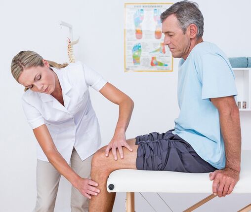
The diagnostic search algorithm is based on considering the nature of the pain syndrome, its duration, identifying symptoms and concomitant events that precede the onset of knee pain. In the first consultation with the physician (traumatologist-orthopedist, surgeon, rheumatologist), visual examination and palpation of the knee are performed, evaluating the volume of active and passive movements. Taking into account the data obtained, in the future, the patient can be assigned:
- laboratory blood tests. . . A complete blood count helps to identify the hematologic changes characteristic of an acute infectious and inflammatory process (leukocytosis, increased ESR), eosinophilia, typical of an allergic reaction. Biochemical and serological studies are more informative for autoimmune diseases, which are characterized by the formation of acute phase proteins and specific immunoglobulins (CRP, rheumatoid factor, ASL-O, SCC, anti-DNA antibodies, etc. ).
- Radiography.The basic diagnostic method is an X-ray of the knee joint in 2 projections. The presence of pathology is indicated by changes in the contours of the head and joint cavity, narrowing of the joint space, changes in the thickness of the endplates, presence of defects in the joint ends of bones, osteolysis and bone destruction. In some diseases (meniscal trauma, Baker's cyst), contrast arthrography demonstrates the greatest sensitivity.
- arthrosonography. . . Knee ultrasound is a quick, inexpensive, accessible, and highly informative diagnostic method. Allows you to assess the presence of effusions and free bodies in the joint cavity, to identify damage and pathological changes in periarticular soft tissues (signs of calcification, hemorrhage, etc. ). They help to differentiate with high precision the etiology of joint pain.
- Tomography and MRI. . . They are the methods of choice for arthropathy of any genesis. They are used for a more detailed assessment of the nature and degree of pathological changes, to identify typical signs of traumatic, inflammatory and tumoral injuries to bone structures and soft tissues. Joint CT and MRI are generally used with limited information content from other instrumental studies.
- joint puncture. . . It is performed when there is an indication of exudate or transudate accumulation in the joint capsule. As part of the differential diagnosis of inflammatory, degenerative and tumoral diseases, a cytological, bacteriological or immunological study of the synovial fluid is carried out. To establish the diagnosis of autoimmune damage to the knee joint, tuberculous arthritis, synovioma, it is extremely important to perform a biopsy of the synovial membrane.
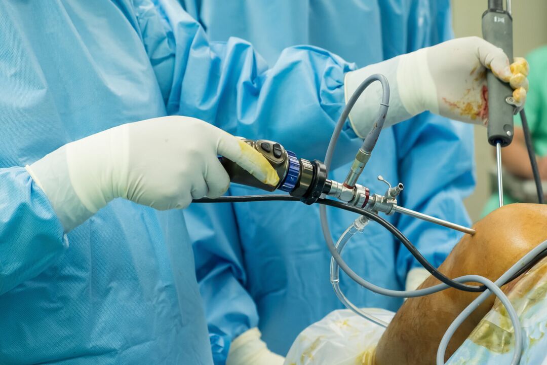
- Arthroscopy. . . The goal of invasive endoscopic diagnosis may be biopsy sampling, elucidating the diagnostic information needed during a visual examination of the joint elements. In some cases, diagnostic arthroscopy evolves into therapeutic (atroscopic removal of intra-articular bodies, meniscectomy, ligament autoplasty, etc. ).
Symptomatic treatment
The treatment of the causes of knee pain is performed in a differentiated manner, taking into account the identified disease. At the same time, symptomatic care is an essential part of a comprehensive treatment process aimed at reducing discomfort and improving quality of life. Immediately after the injury, it is recommended to apply a cold compress to the knee area - this will help reduce pain sensitivity. Ethyl chloride has a local anesthetic and cooling effect. In all cases, resting the knee helps to reduce pain. It is necessary to limit the movement, to give the leg a position where the pain is minimal. When walking, a fixation bandage is applied to the knee, limb immobilization is possible with the aid of a plaster cast.
In the acute period of injury or illness, it is strictly forbidden to massage the knee, apply hot compresses and wear high-heeled shoes. The main classes of medications used for the symptomatic treatment of pain and inflammation are analgesics and NSAIDs in the form of ointments, pills and injections. The measures listed may only temporarily reduce pain, but they do not eliminate the root cause of the arthralgia. Therefore, all cases of knee pain require qualified diagnosis and treatment, and some conditions (fractures, dislocations, hemarthrosis) require emergency medical attention. You cannot postpone a visit to the doctor if the pain is combined with a change in the shape of the knee (swelling, smoothing of contours, asymmetry), inability to perform flexion-extensor movements, patellar ball, impaired limb support.